
Research Areas
Research Areas
Neurovalida scientists have focused on neurodegenerative disorders such as Alzheimer’s disease, Parkinson’s disease, Huntington’s disease and Motor Neurone disease, and they have published 100’s of research papers on these topics in leading international journals. In particular, they have published major reviews and research articles in the following areas:-
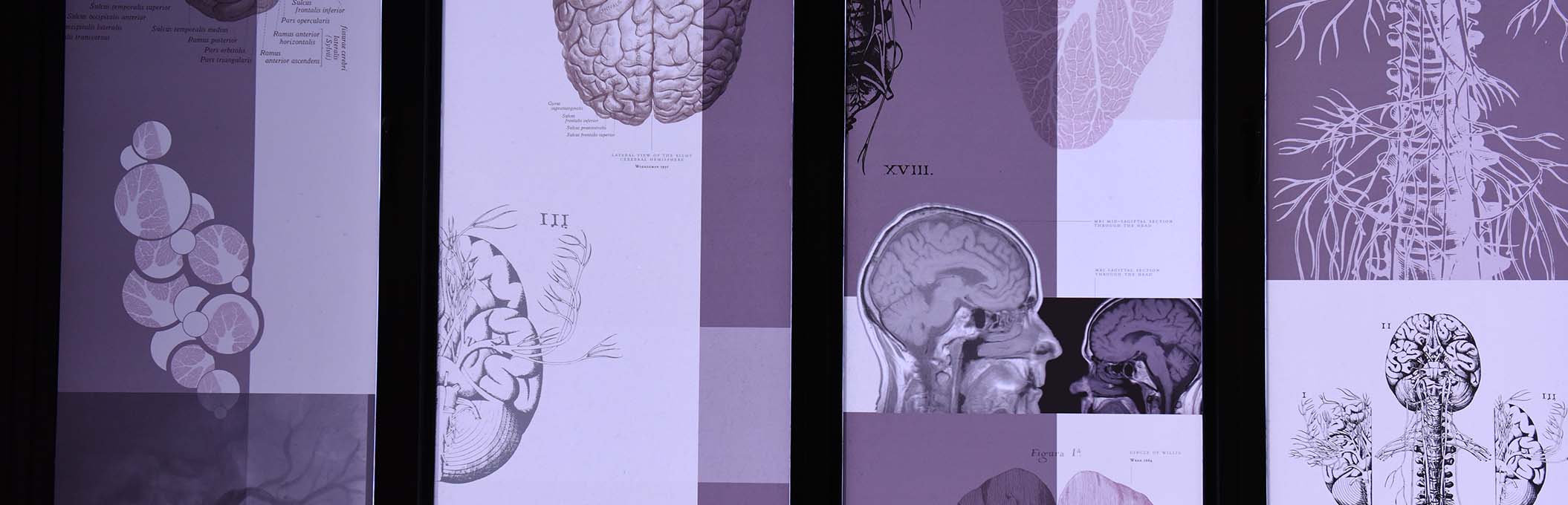
- The human brain in neuroscience research and CNS drug discovery
- Human brain tissue microarray
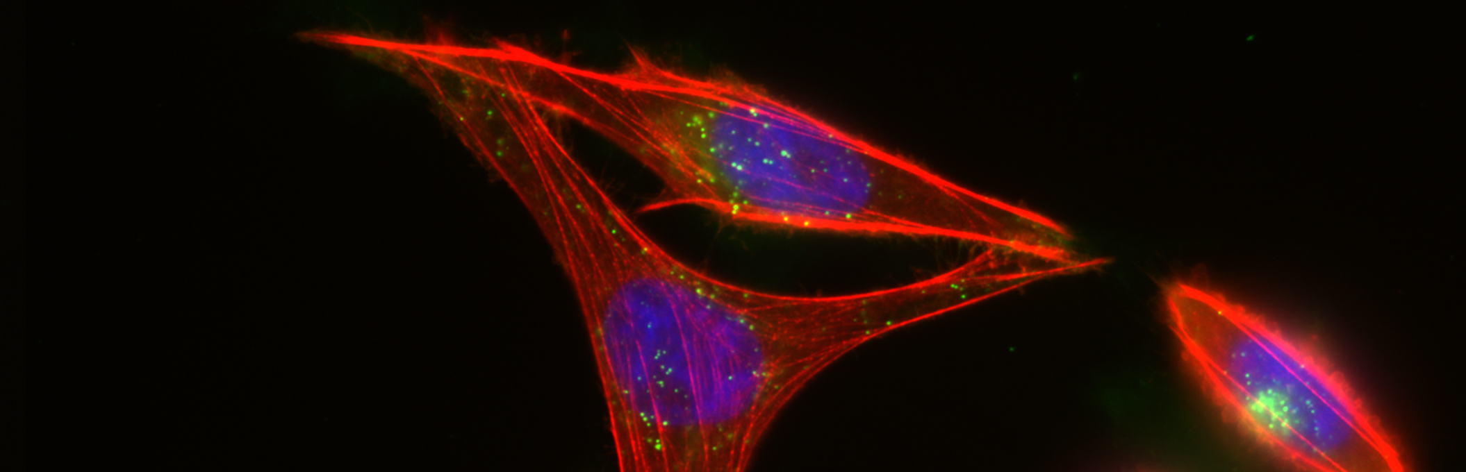
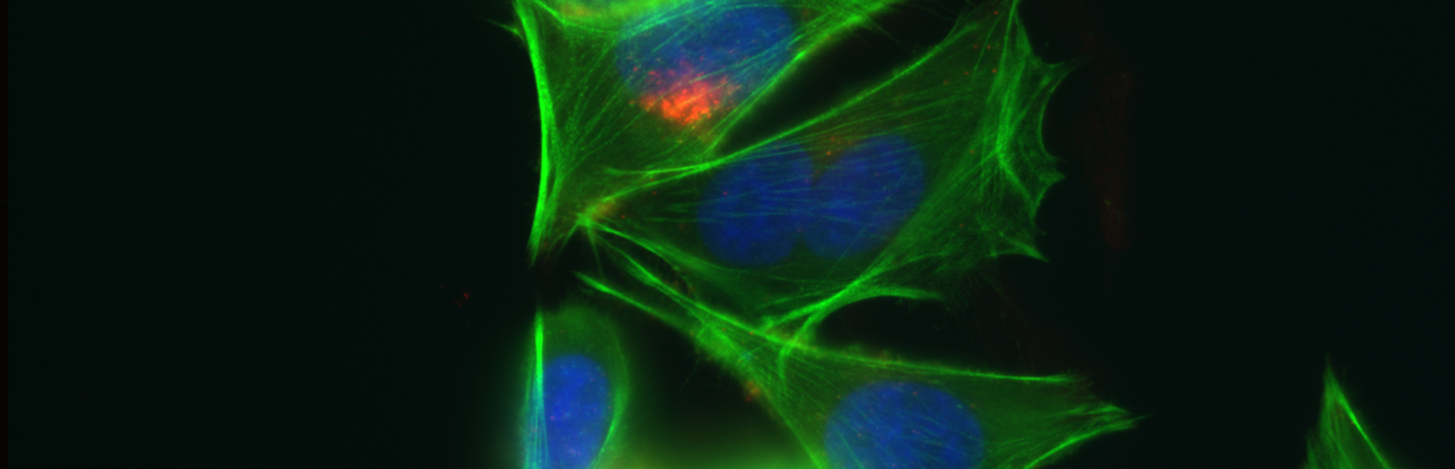
- High content image analysis
- Human receptor autoradiography
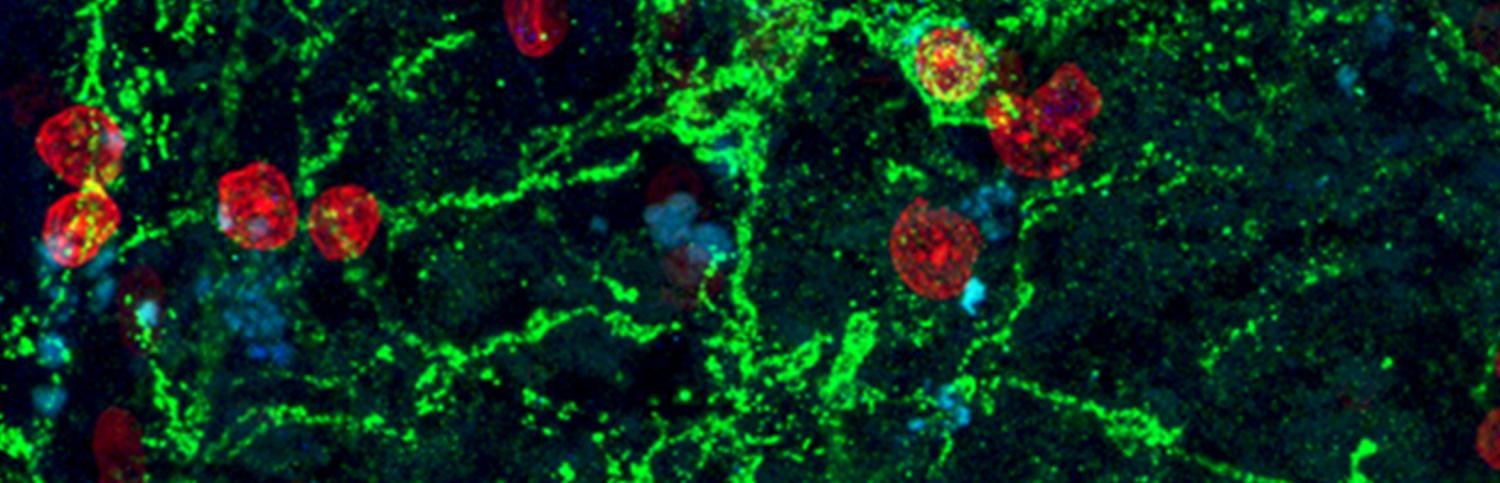
- Human brain immunohistochemistry
- Neuropharmacology/Neurochemistry
Tissue Microarray
Tissue microarray is a method for studying as many as 60 pieces of brain tissue simultaneously. The method works by taking a small sample (like a core biopsy) of brain tissue approximately 2 mm in diameter and setting it into a wax block.
The array can contain either have many different brain regions or cores from many different brains or a combination of the two.
Once the array is made, thin sections (typically 5-10 um in thickness) can be cut from the array using standard histological techniques.
The section containing 60 brain samples then undergoes immunohistochemistry or a similar process. Because all of the brain samples are processed on the same glass microscope slide there is no variation in the processing from one case or region to the next and thus reproducibility is very high.
Once stained the slide can be scanned at high resolution and Metamorph journals can be used to analyse the data.
The analysis can include rich information about the drug target such as cellular and sub-cellular distribution, levels of expression in neurologically normal compared to diseased human brain, and many other features.
Neurovalida scientists then analyse the data and provide a detailed report accompanied by the raw image and image analysis data to the client.
Automated Image Acquisition
We use the VSlide Scanner to acquire images from tissue microarrays and then feed these images into our automated (high content) image analysis program.
Automated (high content) image analysis
We use the Metamorph programme to perform automated high content image analysis which extracts both simple and complex features from the acquired images of the drug target, including cellular and sub-cellular localisation and many other features.
Interpretation of this data is then performed by the Neurovalida research team, who have a combined 60 years of expertise studying the anatomy and chemistry of the human brain.